Good vision supports independence, learning, work and the simple pleasures of daily life. A comprehensive eye exam is more than a quick vision check — it is a deliberate, head-to-toe evaluation of how your eyes are functioning today and how they may change over time. Routine, thorough exams help protect sight and catch emerging problems when they are easiest to manage.
During a comprehensive eye exam we evaluate visual clarity, eye coordination and the health of internal structures. We also screen for common conditions that can progress silently, such as glaucoma, macular degeneration, and diabetic eye disease. Because the eyes often reflect broader health issues, an eye exam can provide early clues about systemic concerns like hypertension or diabetes.
Children, adults, and older adults all benefit from periodic, personalized eye care. For young people, exams confirm visual development and detect learning-related vision problems; for adults, exams preserve productivity and comfort; and for older adults, they help monitor age-related changes that can affect independence and safety.
At Specs Appeal we take time to understand your visual needs and medical history so that every exam delivers both clarity of vision and a clear picture of ocular health.
A complete eye exam looks beyond the simple question of whether you need a new glasses prescription. It examines how your eyes move and work together, inspects the front and back structures of the eye, and evaluates risk factors that could threaten vision in the future. This comprehensive approach helps build a long-term plan for maintaining sight and comfort.
The clinician begins with a conversation about your medical history, medications, family eye health, and any symptoms you may be noticing. Lifestyle details — how much screen time you have, whether you work in visually demanding tasks, or whether you play sports — inform which tests are most relevant and how we interpret their results.
Screening is a major component of every exam. Many sight-threatening conditions progress quietly and can be difficult to notice without careful testing; finding them early gives you many more options for treatment and preservation of vision. A comprehensive exam is the best defense against silent eye disease.
Because the eye provides a unique view of blood vessels and nerve tissue, an attentive eye exam can also flag systemic conditions such as cardiovascular disease, autoimmune disorders, or metabolic disease. These findings are valuable to both eye care providers and your wider healthcare team.

Most comprehensive eye exams take between 30 and 60 minutes, depending on your needs and whether specialized testing is required. Appointments include measured vision testing, evaluation of eye coordination and focusing, and a close inspection of the front and back of the eye using magnified instruments.
Patients can expect a sequence of comfortable, noninvasive tests tailored to their age and medical background. After testing, the clinician reviews findings, answers questions, and outlines any follow-up care or monitoring that may be recommended. This is also an opportunity to discuss lens options, occupational needs, and strategies to reduce eye strain.
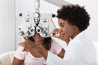
Typical assessments performed during a comprehensive exam include measurements of acuity and color vision, checks of eye alignment and movement, and focused inspections of the cornea, iris, lens and retina. Specialized measurements such as corneal curvature and intraocular pressure are included when indicated.
Below are commonly included tests and what each reveals about your vision and eye health:
Visual acuity testing – Measures how clearly you see at a distance and up close, establishing whether corrective lenses are required and how effective they are.
Color vision testing – Identifies inherited color deficiencies and detects changes in color perception that can signal health issues.
Stereopsis testing – Assesses depth perception and how well both eyes work together to produce a three-dimensional view of the world.
Eye muscle testing – Evaluates the strength and coordination of the muscles that move your eyes to identify strain, misalignment or tracking problems.
Pupil testing – Observes how the pupils respond to light and accommodation, a quick window into neurological and ocular function.
Autorefraction – A computerized scan that provides an initial estimate of refractive error to speed and refine the prescription process.
Retinoscopy – Uses reflected light to help estimate lens power, especially useful for children and nonverbal patients.
Refraction – The clinician’s fine-tuning of the prescription so your lenses deliver the sharpest, most comfortable vision possible.
Keratometry – Measures the curvature of the cornea, information that’s essential for contact lens fittings and some surgical assessments.
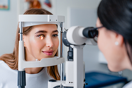
Slit-lamp examination – A magnified inspection of the front and internal structures of the eye to detect inflammation, wear, or disease.
Peripheral visual field – Evaluates side vision and screens for blind spots that can indicate retinal or optic nerve issues.
Intraocular pressure measurement – Screens for elevated pressure, a risk factor for glaucoma that benefits from early detection.
Pupil dilation – Widening the pupils allows a clearer, more complete view of the retina and optic nerve, improving the ability to detect retinal disease and vascular changes.
Please note: dilating eye drops take about 20 minutes to work, and your eyes may be sensitive to light for a few hours following your exam. It’s wise to bring sunglasses to your visit or have someone drive you home from the exam.
When indicated, advanced imaging and diagnostic tests provide high-resolution documentation of retinal structure and function. Examples include optical coherence tomography, fundus photography, corneal topography, and automated visual field testing. These tools allow precise tracking of changes over time and support earlier, more targeted interventions.
At Specs Appeal we pair clinical examination with modern imaging when appropriate so that you leave each visit with a clear understanding of your current eye health and any steps needed to protect it.
Refractive errors occur when the eye does not focus light precisely on the retina, which results in blurred vision at certain distances. They are extremely common and range from nearsightedness to age-related changes in near focus.
Using a camera lens analogy helps make the concept approachable: just as a misfocused camera lens produces a soft image, an eye with refractive error delivers an unfocused retinal image. Symptoms can include blurry vision, headaches from eye strain, difficulty with night driving, or trouble concentrating on small print.
Detecting refractive error is a routine part of the comprehensive exam. Corrections are individualized based on lifestyle, visual demands and long-term eye health considerations.
Myopia (nearsightedness) – Objects in the distance appear blurred because the eye focuses images in front of the retina, often due to an elongated eyeball or steep cornea.
Hyperopia (farsightedness) – Near tasks are more difficult when the eye focuses images behind the retina, which can happen if the eye is shorter than average.
Astigmatism – An irregular corneal or lens shape causes light to bend unevenly, producing distorted or doubled images at any distance.
Presbyopia – A normal, age-related loss of near focusing that typically becomes noticeable in the 40s as the lens becomes less flexible.
Diagnosis begins with visual acuity checks and objective measurements of how light travels through the eye. The refraction portion of the exam refines the prescription so lenses — whether glasses or contact lenses — provide the clearest, most comfortable vision.
Correction options include eyeglasses, a wide range of contact lens materials and designs, or discussions about refractive procedures when appropriate. Each choice has trade-offs related to convenience, visual performance and ocular surface health, and the clinician will help you weigh options based on your individual needs.
Beyond optical correction, management can include targeted strategies for digital eye strain, advice on visual ergonomics, and monitoring plans for conditions that can accompany refractive changes — ensuring your vision solution remains effective as your needs evolve.

Aging brings predictable changes to vision and an increased risk of eye disease. While presbyopia commonly emerges in midlife, other conditions — cataract formation, age-related macular degeneration, glaucoma and vascular eye disease — become more likely with advancing years. Regular exams allow early detection and timely intervention.
A baseline comprehensive exam around age 40 is a practical milestone because many ocular and systemic changes begin to appear at that stage of life. From there, exam frequency is individualized based on findings and health history; some people need annual visits while others require monitoring at specific intervals.
Routine care for older adults also focuses on preserving daily function: optimizing vision for reading, mobility and safe driving, managing dry eye, and coordinating care for systemic diseases that affect ocular health.

Children’s vision develops rapidly during the first years of life, and early exams ensure the visual skills needed for learning and play are on track. Identifying problems such as amblyopia (lazy eye), significant refractive errors, eye alignment issues, or visual processing concerns during infancy and early childhood greatly improves the likelihood of successful treatment.
Guidelines from professional organizations recommend vision assessments in infancy, again in the preschool years, before school entry, and then at regular intervals during childhood. The exact schedule is adapted if a child has risk factors or identified vision concerns.
Child-focused exams use age-appropriate techniques to measure acuity, tracking, focusing and binocularity. When needed, treatment pathways include eyeglasses, patching or vision therapy, and close follow-up — all aimed at giving a child the visual foundation they need to learn and thrive.
Summary: A comprehensive eye exam is a proactive investment in your vision and overall health. From detecting silent disease to tailoring vision solutions for work, school, and life, a thorough exam gives you clear answers and a roadmap for maintaining sight. Contact us for more information or to discuss how often you should schedule an exam.
A comprehensive eye exam is a thorough evaluation of vision and ocular health that goes beyond a basic sight check. It assesses visual clarity, eye alignment and movement, the health of the front and back structures of the eye, and screens for conditions that can progress without obvious symptoms. The goal is to identify current needs and build a plan to preserve comfortable, functional vision over time.
Routine comprehensive exams are valuable at every stage of life because they detect changes early and guide timely care. At Specs Appeal we combine careful clinical testing with a discussion of your medical history and daily visual demands to make each visit meaningful and actionable. Your clinician will review findings, answer questions, and recommend follow-up or monitoring tailored to your situation.
Typical components include visual acuity and refraction to determine prescription needs, checks of eye alignment and binocular function, color vision screening, and measurement of intraocular pressure to assess glaucoma risk. The exam usually incorporates a slit-lamp inspection of the cornea, lens and anterior structures and a detailed view of the retina and optic nerve, often with dilation when indicated. Additional functional assessments may include peripheral visual field testing and stereopsis (depth perception) evaluation.
Objective scans such as autorefraction, retinoscopy and keratometry often support the refraction process and contact lens planning. The clinician selects tests based on age, symptoms and health history so the exam is tailored to what matters most for your eyes. Results are discussed in context, including any recommended imaging, monitoring intervals, or vision solutions.
Most comprehensive eye exams take between 30 and 60 minutes, with the exact length depending on your age, medical history and whether specialized testing or dilation is needed. Pediatric exams, contact lens fittings, or visits that include advanced imaging can extend the appointment time. The clinician schedules enough time to perform tests thoroughly and to review results with you without feeling rushed.
If dilation is part of your visit, plan for additional time because the drops can take about 20 minutes to produce full pupil enlargement and the effects may last several hours. Bringing a list of medications and any current eyewear helps the visit run smoothly. If you wear contact lenses, follow any pre-appointment instructions about removal to ensure accurate measurements.
People of all ages benefit from comprehensive eye exams, but recommended frequency varies by age, risk factors and existing eye conditions. Children should be evaluated at key developmental stages to ensure visual skills for learning are on track, adults generally benefit from periodic exams to maintain comfort and productivity, and a baseline exam around age 40 helps detect early signs of age-related eye disease. Individuals with diabetes, a family history of glaucoma or macular degeneration, or symptoms such as sudden vision changes need more frequent monitoring.
Your clinician will recommend an exam schedule based on findings and health history rather than using a one-size-fits-all timeline. Some patients require annual visits, while others are advised to return at longer intervals with targeted monitoring in between. Consistent follow-up allows timely intervention and better long-term visual outcomes.
Comprehensive exams combine functional testing with structural inspection and, when appropriate, imaging to reveal early signs of disease before symptoms are obvious. Tests such as intraocular pressure measurement, peripheral visual field screening, and detailed retinal evaluation can detect glaucoma, retinal disease, and other conditions in their initial stages. Advanced imaging tools document subtle changes in retinal layers, the optic nerve and corneal shape to track progression over time.
Because many sight-threatening conditions progress without noticeable symptoms, early detection through careful screening substantially increases treatment options and preserves vision. Clinicians also review systemic health factors and medication histories that influence ocular risk and coordinate findings with your broader healthcare team when appropriate. This integrated approach helps protect both eye health and overall well-being.
Pupil dilation involves using eye drops to widen the pupils so the clinician can get an unobstructed, detailed view of the retina and optic nerve. Dilation improves the ability to detect retinal tears, macular changes, vascular abnormalities and other conditions that can be missed through an undilated pupil. It is a safe, routine part of many comprehensive exams and is performed when the clinician determines the benefit outweighs any temporary inconvenience.
After dilation you may experience light sensitivity and blurred near vision for a few hours, so bringing sunglasses and arranging for safe transportation if needed is wise. The drops take effect in roughly 15–30 minutes and wear off gradually; driving immediately afterward can be uncomfortable for some people. Your clinician will explain the reason for dilation and what to expect so you can plan your day accordingly.
Refractive errors such as myopia, hyperopia, astigmatism and presbyopia are identified through objective measurements and a refined refraction process that determines the lens power needed for clear vision. Tests like autorefraction, retinoscopy and clinician-guided refraction work together to produce an accurate prescription for glasses or contact lenses. Symptom review—headaches, eye strain, difficulty reading or trouble with night driving—also informs the assessment.
Correction options include eyeglasses, a variety of contact lens designs and, for appropriate candidates, discussions about refractive procedures. Each option has trade-offs related to comfort, visual needs and ocular surface health, and the clinician helps patients weigh those factors. Follow-up visits ensure prescriptions remain accurate and that any contact lens fit or ocular surface issues are managed effectively.
Advanced imaging tests such as optical coherence tomography (OCT), fundus photography, corneal topography and automated visual field testing provide high-resolution documentation of retinal structure, nerve fiber layers and corneal shape. These tools detect and quantify subtle changes that are not always visible on standard examination and allow precise monitoring of conditions like macular degeneration, glaucoma and corneal disease. Imaging creates a baseline record that improves diagnostic accuracy and helps clinicians spot progression earlier.
When indicated, these tests are integrated with the clinical exam to form a comprehensive picture of ocular health. Results guide treatment decisions and long-term monitoring plans so care can be targeted and timely. Specs Appeal pairs clinical examination with modern imaging when appropriate so patients leave the visit with clear information about their eye health and any next steps.
Children's visual systems develop rapidly, and comprehensive exams identify issues that can interfere with learning and development, such as amblyopia, significant refractive errors, or binocular vision problems. Early detection and treatment of these conditions greatly improve the likelihood of successful outcomes and reduce the risk of long-term deficits. Exams also assess tracking, focusing, and visual processing skills that are essential for reading and classroom performance.
Age-appropriate testing techniques are used so accurate information can be gathered even from very young children, and treatment plans may include eyeglasses, patching or vision therapy when indicated. Regular follow-up ensures progress is tracked and adjustments are made as a child grows. Clinicians communicate findings and recommendations to parents and, when relevant, to educators to support learning-related needs.
To prepare, bring any current eyewear and a list of medications, medical conditions and family eye health history so the clinician has the context needed to interpret test results. If you wear contact lenses, follow any pre-appointment instructions about removal to ensure accurate corneal measurements and refraction. Arrive a few minutes early to complete any intake forms and note any symptoms or questions you want to address during the visit.
During the exam be ready to discuss your daily visual tasks, screen time, and any occupational or recreational demands that affect your eye care needs. Ask about strategies to reduce digital eye strain, whether advanced imaging would be useful, and what follow-up schedule is recommended for your situation. If you prefer to speak with a specific provider, Dr. Jennifer Friehe or another member of the care team will review your results and outline next steps before you leave.
Whether you are updating your glasses, scheduling an eye exam, or exploring contact lenses, Specs Appeal is here to help you see and feel your best.
Contact Us
Learn More
Hours